Jul 18, 2026
Cardiac MRI Scarring Above 5% Predicts Sudden Death Risk
Cardiac MRI Scarring Above 5% Predicts Sudden Death Risk

Updated: Jun 30, 2026, 1:50:24 PM
Text Size:
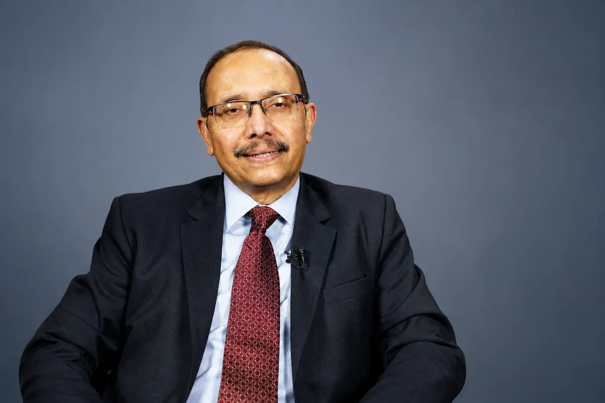
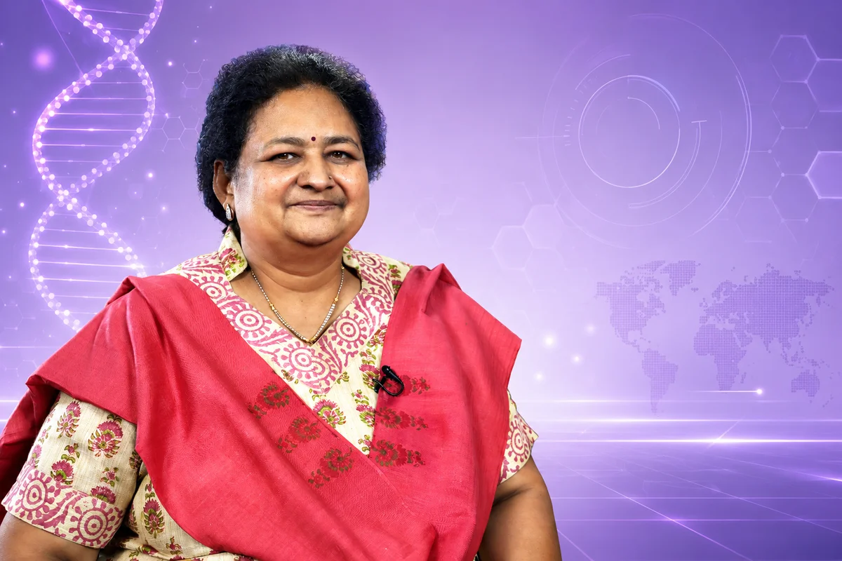
Cardiac MRI Scarring Above 5% Predicts Sudden Death Risk: Dr. Y. S. Chandrashekhar
The clinical reality of managing coronary artery disease (CAD) today often exposes a significant diagnostic gap: the inability of traditional echocardiograms or nuclear perfusion mismatch techniques to precisely distinguish between hibernating and dead myocardium.
Unresolved diagnostic ambiguities frequently lead to sub-optimal revascularization planning, leaving complex cardio-kidney-metabolic (CKM) patients vulnerable to unaddressed ventricular arrhythmias and sudden cardiac death.
In an interview at TheRightDoctors studio at the 4th World Congress on Cardio-Kidney-Metabolic Medicine (WCCKMM 2026) at the Leela, Mumbai, Dr. P. C. Manoria, Executive President of the congress, spoke with Dr. Y. S. Chandrashekhar, Editor-in-Chief, JACC Cardiovascular Imaging (USA), on advancing myocardial viability assessment.
Probing the viability practice gap, Dr. P. C. Manoria asked which imaging modality offers the most reliable diagnostic data. Dr. Y. S. Chandrashekhar stated that late gadolinium enhancement (LGE) in cardiac MRI now defeats alternative techniques by turning dead myocardium white while normal tissue remains black.
Responding to Dr. P. C. Manoria’s query about quantifying arrhythmic propensity, Dr. Y. S. Chandrashekhar noted that scar size is highly proportionate to future adverse events. In ischemic cardiomyopathy, LGE involving over 5% of the myocardium identifies a high-risk group.
Dr. Y. S. Chandrashekhar highlighted that this threshold preserves significant prognostic power even in patients presenting with mild to moderately reduced ejection fractions, resolving clinical dilemmas regarding implantable cardioverter-defibrillator (ICD) placement.
Elaborating on structural scar patterns as prompted by Dr. P. C. Manoria, Dr. Y. S. Chandrashekhar explained that sub-endocardial scars following a specific coronary artery territory guarantee an ischemic origin, even if spontaneous recanalization has occurred.
When Dr. P. C. Manoria shifted focus to the microvasculature, Dr. Y. S. Chandrashekhar acknowledged that while epicardial stenosis is simpler to mechanically resolve, microvascular dysfunction requires sophisticated non-invasive quantification via PET or invasive index of microvascular resistance (IMR).
Raised by Dr. P. C. Manoria as a critical closing provocation: Is the clinical community ready to move past the legacy of ejection fraction and adopt global longitudinal strain as the primary measure of contractility, or will established guidelines continue to anchor practice to traditional metrics?
TheRightDoctors | Official Digital Knowledge Partner | WCCKMM 2026
ALSO READ | Dr Chandrashekhar: Image Fat Depot Phenotypes | WCCKMM 2026
ALSO WATCH | The Indispensable Role of SGLT2i in Heart Failure
Support TheRightDoctors
A fake news pandemic has taken the world by storm. More so for medical content.
We are witnessing a paradox of information overload and unanswered questions among both the lay public and the medical community.
TheRightDoctors brings you Insights from thought leaders across leading medical specialities. Views which help correct the information asymmetry and articulate the voice of medical science and reason.
This effort, however, needs your support.
Medical journalism is a niche which can survive only when it is adequately funded.
If you appreciate the work we do, we would like to request your support. Every contribution however big or small is valuable to us.
The options on this page enable you to choose between a one-time, monthly or annual payment. This is inclusive of GST. If you are a firm with GST, we will issue a GST invoice. Please reach out to us at support@therightdoctors.com
We thank you for your trust and your support.
SupportTags: Dryschandrashekhar | Drpcmanoria | Wcckmm2026 | Therightdoctors | Cardiology | Cardiacmri | Jaccimaging | Cardiokidneymetabolic | Ckm | Myocardialviability | Suddencardiacdeath | Ischemiccardiomyopathy | Microvasculardysfunction | Globallongitudinalstrain | Ejectionfraction | Medicalimaging | Mumbaiconference | Cardiometabolicmedicine |



Most Popular

Dr. Pankaj Manoria on Post-MI Antiplatelet Therapy Selection

Understanding Systemic Burnout and Policy Initiatives for Doctors

Beyond the textbook: Overhauling Medical Curricula

Eradicating Administrative Burden on National Doctors Day
More from TheRightDoctors

Food Sequencing & Unpolished Grains Beat Sugar Substitution in CKM
Umbilical Cord LDL of 25 mg/dL is the True Standard: Dr Rajeev Agarwal
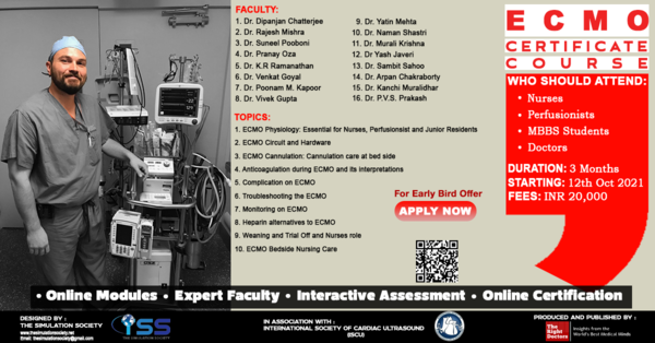
Click here to Register for ECMO course
Trending
Carb Quality and Portion Control Outweigh Elimination: Dr. Niti Desai
UACR Above 10 mg/g Signals Vascular Damage: Dr. Anil Pareek
Sweeteners Disrupt Insulin Secretion, Warns Dr Shaifali Bansal

Pregnancy Programming Dictates Adult Metabolic Risk: Dr. Sunil Gupta
Powered by:
.png)
Awards & Recognitions:


